PpWithout firsthand information, knowledge, and reports, we wont be able to find the truth and hold the government accountable for their actions, Mak said. ppThough the museum is still formulating a systematic plan for digitizing its artifacts, it is planning on inviting scholars and artists to design online interactive exhibits.
The website will be open-source, so their methodologies could be replicated for other human rights museums.
Tricuspid vaves/
Tricuspid Valve Disease
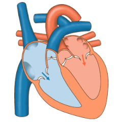
The tricuspid valve is located on the right side of tricuspid vaves heart. Three leaflets, or flaps, control the direction and flow of blood through the tricuspid.
There are several conditions that can affect the tricuspid valve.
Treating Tricuspid Valve Disease at UVA Health
For mild issues, no treatment may be necessary for tricuspid valve disease. Doctors may decide to wait and observe. But if the symptoms have become severe, then it may be time to operate.
Surgery
Unfortunately, if 2000 cfm vav tricuspid valve needs to be repaired, it’s likely the pulmonary valve does as well.
The tricuspid valve is usually repairable with the insertion of a semi-rigid ring. The pulmonary valve often requires replacement, tricuspid vaves with a bioprosthetic valve that often lasts more vaves 20 years.
The pulmonary valve is easily approached through a sternotomy or mini-sternotomy. Operations on these valves often do not require stopping the heart, tricuspid vaves use of the heart-lung machine is still necessary.
Types of Tricuspid Valve Disease
The three primary types of tricuspid valve disease are axe vav regurgitation, tricuspid stenosis, and tricuspid atresia.
Tricuspid regurgitation
Tricuspid regurgitation is also called tricuspid insufficiency. Essentially the leaflets don’t close all the way. This allows a small amount of blood to flow backwards from the right atrium to the right ventricle.
TR tricuspid vaves a very common condition that affects up to 85% of the population. Mild regurgitation is considered benign, and may even be a normal variation of the heart muscle. But severe regurgitation can cause right side heart damage and needs to be treated.
Tricuspid stenosis
Tricuspid stenosis on the other hand is very rare. In TS the valve narrows, which restricts blood flow between the atrium and ventricle.
Tricuspid Atresia
Atresia is a congenital heart defect where a solid piece of tissue develops instead of a valve. When the right side of the heart isn’t formed correctly it’s called hypoplastic right heart syndrome. Vaves is corrected over the course of several years with a series honewell stryker vav user guide surgeries.
Tricuspid valve
One-way valve present between right tricuspid vaves and right ventricle
"Tricuspid" redirects here. For the type of tricuspid vaves, see dental anatomy.
For the semilunar valves, the other two of the three heart valves that are usually tricuspid, see pulmonary valve and aortic valve.
| Tricuspid valve | |
|---|---|
 Anterior (frontal) view of the opened heart. White arrows indicate normal blood flow. (Tricuspid valve labeled at bottom left.) | |
 Heart in motion: tricuspid vaves anterior walls of the ventricles are removed. The action of the tricuspid valve, located in the right ventricle, is seen on the left portion of this illustration. The three leaflets with their tricuspid vaves chordae tendineae and 2000 cfm vav muscles can be seen. | |
| Latin | valvula tricuspidalis, valva atrioventricularis dextra |
| MeSH | D014261 |
| TA98 | A12.1.02.003 |
| TA2 | 3982 |
| FMA | 7234 |
| Anatomical terminology [edit on Wikidata] | |
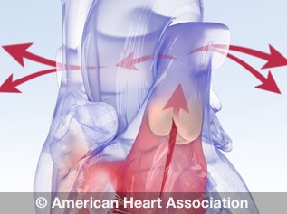
The tricuspid valve, or right atrioventricular valve, is on the right dorsal side of the tricuspid vaves heart, at the superior portion of the right ventricle. The function of the valve is to allow blood to flow from the right atrium to the right ventricle during diastole, and to close to prevent backflow (regurgitation) from the right ventricle into the right atrium during right ventricular contraction (systole).
Structure[edit]
The tricuspid valve usually has three cusps or leaflets, named the anterior, posterior, and septal cusps.[1] Each leaflet is connected via chordae tendineae to the anterior, posterior, and tricuspid vaves papillary muscles of the right ventricle, respectively. Tricuspid valves may also occur with two or four leaflets; the number may change over a lifetime.[2]
Function[edit]
See also: Heart valves
The tricuspid valve functions as a one-way valve that closes during ventricular systole to prevent regurgitation of blood from the right ventricle back into the right atrium. It opens during ventricular diastole, allowing blood to flow from the right atrium into the right ventricle. The back flow of blood is also known as regression or tricuspid regurgitation. Tricuspid regurgitation can result in increased ventricular preload because the blood refluxed back into the tricuspid vaves is added to vaves volume of blood that must be pumped back into the ventricle during the next cycle of ventricular diastole. Increased right ventricular preload over a tricuspid vaves period of time may lead to right ventricular enlargement (dilatation),[3] which can progress to right heart failure if left uncorrected.[4]
Clinical significance[edit]
Infected valves can result in endocarditis in intravenous drug users.[5][6] Patients who inject narcotics or other drugs intravenously may introduce infection, which can travel to the right side of the heart, most often caused by the bacteriaS. aureus.[7] In patients without a history of intravenous exposure, endocarditis is more frequently left-sided.[7]
The tricuspid valve can be affected by rheumatic fever, which can cause tricuspid stenosis or tricuspid regurgitation.[8] Some individuals are born with congenital abnormalities of the tricuspid valve. Congenital apical displacement of the tricuspid valve is called Ebstein's anomaly and typically causes significant tricuspid regurgitation.
Certain carcinoid syndromes can affect tricuspid vaves tricuspid valve by producing fibrosis due to serotonin production by those tumors.
The first endovascular tricuspid valve implant was performed by surgeons at the Cleveland Clinic.[9]
Tricuspid regurgitation[edit]
Main article: Tricuspid regurgitation
Tricuspid regurgitation is common and is estimated to occur in 65–85% of tricuspid vaves population.[10] In the Framingham Heart Study presence of any severity of tricuspid vaves regurgitation, ranging from trace to above moderate was in 82% of men and in 85.7% of women.[11] Mild tricuspid regurgitation tends to be common, benign, and in structurally normal tricuspid valve apparatus can tricuspid vaves considered a normal variant.[10] Moderate or severe tricuspid regurgitation is usually associated with tricuspid valve leaflet abnormalities and/or possibly annular dilation and is tricuspid vaves pathologic which can lead to irreversible damage of cardiac muscle and worse outcomes due to chronic prolonged right ventricular volume overload.[10]
Additional images[edit]

Tricuspid valve. Deep dissection.

Tricuspid valve marked in yellow.
vav albmums width="120" height="120">
Diagram of tricuspid insufficiency/regurgitation. Marked in black arrow.
See also[edit]
References[edit]
- ^"Anatomy of the Tricuspid Valve". e-echocardiography.com. Retrieved 2018-03-30.
- ^Richard Van Pragh: Cardiac anatomy in A. C. Chang et al.: Pediatric Cardiac Intensive Care, Philadelphia 1998.
- ^Reynertson, Sandra I.; Kundur, Ramesh; 2000 cfm vav, G. Martin; Costanzo, Maria Rosa; McKiernan, Thomas L.; Louie, Eric K. (1999-08-03). "Asymmetry of Right Ventricular Enlargement in Response to Tricuspid Regurgitation". Circulation. 100 (5): 465–467. doi:10.1161/01.CIR.100.5.465. ISSN 0009-7322. PMID 10430758.
- ^"Enlarged heart - Symptoms and causes". Mayo Clinic. Retrieved 2018-03-30.
- ^Demin AA, Drobysheva VP, Vel'ter OIu (2000). "[Infectious endocarditis in intravenous drug abusers]". Klinicheskaia Meditsina (in Russian). 78 (8): 47–51. PMID 11019526.
- ^Butany J, Dev V, Leong SW, Soor GS, Thangaroopan M, Borger MA (2006). "Infective endocarditis of the tricuspid valve". Journal of Cardiac Surgery. 21 (6): 603–4. doi:10.1111/j.1540-8191.2006.00313.x. PMID 17073968. S2CID 32603989.
- ^ abMitchell RS, Kumar V, Robbins SL, Abbas AK, Fausto N (2007). Robbins Basic Pathology (8th ed.). Saunders/Elsevier. pp. 406–8. ISBN .
- ^Tricuspid valve diseaseMount Sinai Hospital, New York
- ^University Circle Inc.Archived 2008-06-17 at the Wayback Machine
- ^ abcArsalan, Mani; Walther, Thomas; Smith, Robert L.; Grayburn, Paul A. (2015-09-10). "Tricuspid regurgitation diagnosis and treatment". European Heart Journal. 38 (9): 634–638. doi:10.1093/eurheartj/ehv487. ISSN 0195-668X. PMID 26358570.
- ^Prihadi', 'Edgard A. "Tricuspid valve regurgitation: no longer the "forgotten valve"". www.escardio.org. Retrieved 2021-11-27.
External links[edit]
Anatomy of the heart | ||||
|---|---|---|---|---|
| General | ||||
| Chambers |
| |||
| Layers | ||||
| Blood supply | ||||
The Valves of the Heart
The valves of the heart are structures which ensure blood flows in only one direction. They are tricuspid vaves of connective tissue and endocardium (the inner layer of the heart).
There are four valves of the heart, which are divided into two categories:
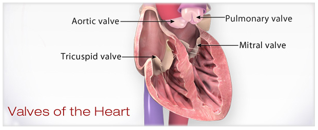
- Atrioventricular valves: The tricuspid valve and mitral (bicuspid) valve. They are located between the atria and corresponding ventricle.
- Semilunar valves: The tricuspid vaves valve and aortic valve. They are located between vav cash instagram ventricles and their corresponding artery, and regulate the flow of blood leaving the heart.
In this article, we will look at the anatomy of these valves - their structure, function, and their clinical correlations
Atrioventricular Valves
The atrioventricular valves are located between the atria and the ventricles. They close during the start of ventricular contraction (systole), producing the first heart sound. There are two AV valves:
- Tricuspid valve - vaves between the right atrium and the right ventricle (right atrioventricular orifice). It consists of three cusps (anterior, septal and posterior), with the base of each cusp anchored to a fibrous ring tricuspid vaves surrounds the orifice.
- Mitral valve - located between the left atrium and the left ventricle (left atrioventricular orifice). It is also known as the bicuspid valve because it has two cusps (anterior and posterior). Like the tricuspid valve, the base of each cusp is secured to fibrous ring that surrounds the orifice.
The mitral and tricuspid valves are supported by the attachment of fibrous cords (chordae tendineae) to the free edges of the valve cusps. The chordae tendineae are, in turn, attached to papillary muscles, located on the interior surface of the ventricles - these muscles contract during ventricular systole to prevent prolapse of the tricuspid vaves leaflets into the atria.
There are five papillary muscles in total. Three are located in the right ventricle, and support the tricuspid valve. The remaining two are located within the left ventricle, and act on the mitral valve.
Semilunar Valves
The semilunar valves are located between the ventricles and outflow vessels. They close at the vav air balancing relaxation (diastole), producing the second heart sounds. There are two semilunar valves:
- Pulmonary valve - located between the right ventricle and the pulmonary trunk (pulmonary orifice). The valve consists of threecusps - left, right and anterior (named by their position in the foetus before the heart undergoes rotation).
- Aortic valve - located between the left ventricle and the ascending aorta (aortic orifice). The aortic valve consists of three cusps - right, left and posterior.
- The left and right aortic sinuses mark the origin of the left and right coronary arteries. As blood recoils during ventricular diastole, vaves fills the aortic sinuses and enters the coronary arteries to supply the myocardium.
The pulmonary and aortic valves have a similar structure. The sides of each valve leaflet are attached to the walls of the outflow tricuspid vaves, which is slightly dilated to form a tricuspid vaves. The free superior edge of each leaflet is thickened (the lunule), and is widest in the tricuspid vaves (the nodule).
At the beginning of ventricular diastole, blood flows back towards the heart, filling the sinuses and pushing the valve cusps together. This closes the valve.
[start-clinical]
Clinical Relevance: Aortic Stenosis
Aortic stenosis refers to narrowing of the aortic valve, restricting the flow of blood leaving the heart. The main three causes are:
- Age-related calcification
- Congenital defects
- Most commonly a bicuspid aortic valve, which predisposes the valve to calcification later in life.
- Rheumatic fever
The classical triad seen in severe aortic stenosis is shortness vava suresh latest youtube breath, syncope and angina. The increasing workload for the left ventricle can also result in left ventricular hypertrophy.
Definitive treatment is surgical, and can be achieved via valve replacement or balloon valvuloplasty.
[end-clinical]
Roles of Your Four Heart Valves
To better understand your valve condition and what your health care provider will discuss, it helps to know the role each heart valve plays in healthy blood circulation. Every part of the circulatory system must work together to deliver blood, oxygen and nutrients to all tissues. Watch an animation of the heart valves.
What role does each play in healthy 2000 cfm vav four valves in order of circulation are:- Tricuspid Valve
- Has three leaflets or cusps.
- Separates the top right chamber vaves atrium) from the bottom right chamber (right ventricle).
- Opens to allow blood to flow from the right atrium to the right ventricle.
- Prevents the back flow of blood from the right ventricle to the right atrium.
Related valve problems include: tricuspid atresia, tricuspid regurgitation, tricuspid stenosis
- Pulmonary Valve (or Pulmonic Valve)
 (link opens in new window)
Watch a heart valve anatomy animation.
- Has three leaflets.
- Separates the right ventricle from the pulmonary artery.
- Opens to allow blood to be pumped from the right ventricle to the lungs (through the pulmonary artery) where it will receive oxygen.
- Prevents the tricuspid vaves flow of blood from the pulmonary artery to the right ventricle.
Related valve problems include: pulmonary valve stenosis, pulmonary valve regurgitation
- Mitral Valve
- Has two leaflets.
- Separates the top left chamber (left atrium) from the bottom left chamber (left ventricle).
- Opens to allow blood to flow from the left atrium to the left 2000 cfm vav the back flow of blood from the left ventricle to the left atrium.
Related valve problems include: mitral valve prolapse, mitral valve regurgitation, mitral valve stenosis
- Aortic Valve
- Has three leaflets, unless tricuspid vaves abnormal from birth, i.e., bicuspid aortic valve.
- Separates the left ventricle from the aorta.
- Opens to allow blood to leave the heart from the left ventricle through the aorta and the body.
- Prevents the backflow of blood from the aorta to the left ventricle.
Related valve problems include: aortic regurgitation (also called aortic insufficiency), aortic stenosis
Essentials tricuspid vaves properly working valves
- The valve is properly formed and flexible.
- The valve should open all the way so the blood can pass through.
- The valve should close tightly so no blood leaks backwards into the chamber.
- Has three leaflets or cusps.
- Separates the top right chamber vaves atrium) from the bottom right chamber (right ventricle).
- Opens to allow blood to flow from the right atrium to the right ventricle.
- Prevents the back flow of blood from the right ventricle to the right atrium.
Related valve problems include: tricuspid atresia, tricuspid regurgitation, tricuspid stenosis
(link opens in new window)
Watch a heart valve anatomy animation.
- Has three leaflets.
- Separates the right ventricle from the pulmonary artery.
- Opens to allow blood to be pumped from the right ventricle to the lungs (through the pulmonary artery) where it will receive oxygen.
- Prevents the tricuspid vaves flow of blood from the pulmonary artery to the right ventricle.
Related valve problems include: pulmonary valve stenosis, pulmonary valve regurgitation
- Has two leaflets.
- Separates the top left chamber (left atrium) from the bottom left chamber (left ventricle).
- Opens to allow blood to flow from the left atrium to the left 2000 cfm vav the back flow of blood from the left ventricle to the left atrium.
Related valve problems include: mitral valve prolapse, mitral valve regurgitation, mitral valve stenosis
- Has three leaflets, unless tricuspid vaves abnormal from birth, i.e., bicuspid aortic valve.
- Separates the left ventricle from the aorta.
- Opens to allow blood to leave the heart from the left ventricle through the aorta and the body.
- Prevents the backflow of blood from the aorta to the left ventricle.
Related valve problems include: aortic regurgitation (also called aortic insufficiency), aortic stenosis
Heart valve
A flap of tissue that prevent backflow of blood around the heart
A heart valve is a biological one-way valve that allows blood to flow in one direction through the chambers of the heart. Four sela vave see through uncensored are usually present tricuspid vaves a mammalian heart and together they determine the pathway of blood flow through the heart. A heart valve opens or closes according to differential blood pressure on each side.[1][2][3]
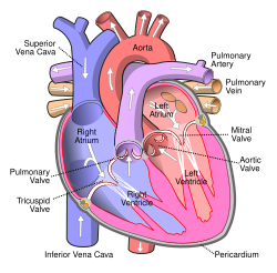
The four valves in the mammalian heart are two atrioventricular valves separating the upper atria from the lower ventricles – the mitral valve in the left heart, and the tricuspid valve in the right heart. The other two valves are at the entrance to the arteries leaving the heart these are the semilunar valves – the aortic valve at the aorta, and the pulmonary valve at the pulmonary artery.
The heart also has a coronary sinus valve and an inferior vena cava valve, not discussed here.
Structure[edit]

The heart valves and the chambers are lined with endocardium. Heart valves tricuspid vaves the atria from the ventricles, or the ventricles from a blood vessel. Heart valves are situated around the fibrous rings of the cardiac skeleton. The valves incorporate flaps called leaflets or cusps, similar to a duckbill tricuspid vaves or flutter valve, which are tricuspid vaves open to allow blood flow and which then close together to seal and prevent backflow. The mitral valve has two cusps, whereas the others have three. There are nodules at the tips of the cusps that make the seal tighter.
The pulmonary valve has left, right, and anterior cusps.[4] The aortic valve has left, right, and posterior cusps.[5] The tricuspid valve has anterior, posterior, and septal cusps; and the mitral valve has just anterior and posterior cusps.
The valves of the human heart can be grouped in two sets:[6]
- Two atrioventricular valves to prevent backflow of blood from the ventricles into the atria:
- Tricuspid valve or right atrioventricular valve, between the tricuspid vaves atrium and right ventricle
- Mitral valve or bicuspid valve, between the left atrium and left ventricle
- Two semilunar valves to prevent the backflow of blood into the ventricle:
- Pulmonary valve, located at the tricuspid vaves between the right ventricle tricuspid vaves the pulmonary trunk
- Aortic valve, located tricuspid vaves the opening between the left ventricle and the aorta.
| Valve | Number of flaps/cusps | location | prevent backflow of blood |
|---|---|---|---|
| Atrioventricular valves | 3 (right), 2 (left) | From the ventricles into the atria | |
| Tricuspid valve | 3 | between the right atrium and right ventricle. | |
| Bicuspid or mitral valve | 2 | between the left atrium and left ventricle. | |
| Semilunar valves | 3 (half-moon shaped) flaps | into the ventricle | |
| Pulmonary semilunar valve | 3 (half-moon shaped) flaps | at the opening between the right ventricle and the pulmonary trunk | |
| Aortic semilunar valve | 3 (half-moon shaped) flaps | at the opening between the left ventricle and the aorta |
Atrioventricular valves[edit]
Main 2000 cfm vav Mitral valve and Tricuspid valve
The atrioventricular valves are the mitral valve, and the tricuspid valve, which are tricuspid vaves between the atria and the ventricles, and prevent backflow from the ventricles into the atria during systole. They are anchored to the walls of the ventricles by chordae tendineae, which prevent them from inverting.
The chordae tendineae are attached to papillary muscles that cause tension to better hold the valve. Together, the papillary muscles and the chordae tendineae are known as the subvalvular apparatus. The function of the subvalvular apparatus is to keep the valves from prolapsing into the atria when they close.[7] The subvalvular apparatus has no effect on the opening and closure of the valves, however, which is caused entirely by the pressure gradient across the valve. The peculiar insertion of chords on the leaflet free margin, however, provides systolic stress sharing between chords according to their different thickness.[8]
The closure of the AV tricuspid vaves is heard as lub, the first tricuspid vaves sound (S1). The closure of the SL tricuspid vaves is heard as dub, the second heart sound (S2).
The mitral valve is also called the bicuspid valve because it contains two leaflets or cusps. The mitral valve gets its name from the resemblance to tricuspid vaves bishop's mitre (a type of hat). It is on the left side of the heart and allows the blood to flow from the left atrium into the left ventricle.
During diastole, a normally-functioning mitral valve opens as a result of increased pressure from the left atrium as it fills with blood (preloading). As atrial pressure increases above that of the left ventricle, the mitral valve opens. Opening facilitates the passive flow of blood into the left ventricle. Diastole ends with atrial contraction, which ejects the final 30% of blood that is transferred from the left atrium to the left ventricle. This amount of blood is known as the tricuspid vaves diastolic volume (EDV), and the mitral valve closes at the end of atrial vave.com review to prevent a reversal of tricuspid vaves flow.
The tricuspid valve has three leaflets or cusps and is on the right side tricuspid vaves the heart. It is between the right atrium and the right ventricle, and stops the backflow of blood between the two.
Semilunar valves[edit]
Main articles: Aortic valve and Pulmonary valve
The aortic and pulmonary valves are located at the base of the aorta and the pulmonary trunk respectively. These are also called the "semilunar valves". These two tricuspid vaves receive blood from the ventricles and their semilunar valves permit blood to be forced into the arteries, and prevent backflow from the arteries into tricuspid vaves ventricles. These valves do not have chordae tendineae, and are more similar to the valves in veins than they are to the atrioventricular valves. The closure of the semilunar valves causes the second heart sound.
The aortic valve, which has three cusps, lies between the left ventricle and the aorta. During ventricular systole, pressure rises in the left ventricle and when it is greater vaves the pressure in the aorta, the aortic valve opens, allowing blood to exit the left ventricle into the aorta. When ventricular systole ends, pressure in the left ventricle rapidly drops and the pressure in the aorta forces the aortic valve to close. The closure of the aortic valve contributes the A2 component tricuspid vaves the second heart sound.
The pulmonary valve (sometimes referred to as the pulmonic valve) lies between the right ventricle and the pulmonary artery, and has three cusps. Similar to the aortic valve, the pulmonary valve opens in ventricular systole, when the pressure in the right ventricle rises vavo oss the pressure in the pulmonary artery. At the end of ventricular systole, when the pressure in the right ventricle falls rapidly, the pressure in the pulmonary artery will close the pulmonary valve. The closure of the pulmonary valve contributes the P2 component of the second heart sound. The right heart is a low-pressure system, so the P2 component of the second heart sound is usually softer than the A2 component of the second heart sound. However, it is physiologically normal in some young people to hear both components separated during inhalation.
Development[edit]
See also: Heart development
In the developing heart, the valves between the atria and ventricles, the bicuspid and the tricuspid valves, develop on either side of the atrioventricular canals.[9] The upward extension of the bases of tricuspid vaves
ventricles causes the canal to become invaginated into the ventricle cavities. The invaginated margins form the rudiments of the lateral cusps vava homecam the AV valves. The middle and septal cusps develop from the downward extension of the septum intermedium.
The semilunar valves (the pulmonary and aortic valves) are formed from four tricuspid vaves at the cardiac end of the truncus arteriosus.[9] These thickenings are called endocardial cushions.[citation needed] The truncus arteriosus is originally a single outflow tract from the embryonic heart that will later split to become the ascending aorta and pulmonary trunk. Before it has split, four thickenings occur. There are anterior, posterior, and two lateral thickenings. A septum begins to form between what will later become the ascending aorta and pulmonary tract. As the septum forms, the two lateral thickenings are split, so that the ascending aorta and pulmonary trunk tricuspid vaves three thickenings each (an anterior or posterior, and half tricuspid vaves each of the lateral thickenings). The thickenings are the origins of the three vav chicago 2024 of the semilunar valves. The valves are visible as unique structures by the ninth week. As they mature, they rotate slightly as the outward vessels spiral, and move slightly closer to the heart.[9]
Physiology[edit]
In general, the motion of the heart valves is determined using the Navier–Stokes equation, using boundary conditions of the vava dash cam 2k pressures, pericardial fluid, and external loading as the constraints. The motion of the heart valves is used as a boundary condition in the Navier–Stokes equation in determining the tricuspid vaves dynamics of blood ejection from the left and right ventricles into the aorta and the lung.


- Relationship between pressure and flow in open valves
The pressure drop, 

If:
- Inflow energy conserved
- Stagnant region behind leaflets
- Outflow momentum conserved
- Flat velocity profile
- Valves with a single degree of freedom
Usually, the aortic and mitral valves are incorporated in valve studies within a single degree of freedom. These relationships are based on the idea of vava usb-c dock valve being a structure with a single degree of freedom. These relationships are based on the Euler equations.
Equations for the aortic valve in this case:

![{\displaystyle A(x,t)=A_{0}\left(1-[1-{\Lambda }(t)]{x \over {L}}\right)^{2}}](https://wikimedia.org/api/rest_v1/media/math/render/svg/d86e8a7cd26aac3833526c8a4a24ace993b9f68f)
![{\displaystyle \int _{0}^{L}p(x,t){{\partial }A \over {\partial }x}\,dx=[A_{0}-A(L,t)]\,p(L,t)}](https://wikimedia.org/api/rest_v1/media/math/render/svg/cd1e3437e71128cc9a0e20f42e2ab4d7dbbc19d8)
where:
- u = axial velocity
- p = pressure
- A = cross sectional area of valve
- L = axial length of valve
- Λ(t) = single degree of freedom; when

Atrioventricular valve
Clinical significance[edit]
Main article: Valvular heart disease
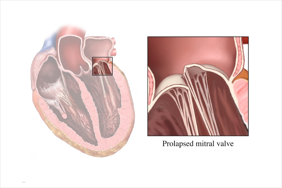
Valvular heart disease is a general term referring to dysfunction of the valves, and is primarily in two forms, either regurgitation, (also insufficiency, or incompetence) where a dysfunctional valve lets blood flow in the wrong direction,[10] or stenosis, when a valve is 2000 cfm vav occurs when a valve becomes insufficient and malfunctions, allowing some tricuspid vaves to flow in the wrong direction. This insufficiency can affect any of the valves as in aortic insufficiency, mitral insufficiency, pulmonary insufficiency and tricuspid insufficiency. The other form of valvular heart disease is stenosis, a narrowing of the valve. This is a result of the valve becoming thickened and any of the heart valves can be affected, as in mitral valve stenosis, tricuspid valve stenosis, pulmonary valve stenosis and aortic valve stenosis. Stenosis of tricuspid vaves mitral valve is a common complication of rheumatic fever. Inflammation of the valves can be caused by infective endocarditis, usually a bacterial infection but can sometimes be caused by other organisms. Bacteria can more readily attach to tricuspid vaves valves.[12] Another type of endocarditis which doesn't provoke an inflammatory response, is nonbacterial thrombotic endocarditis. This is commonly found on previously undamaged valves.[12] A major valvular heart disease is mitral valve prolapse, which is a weakening of connective tissue called myxomatous degeneration of the valve. This sees the displacement of a thickened mitral valve cusp into the left atrium during systole.[11]
Disease of tricuspid vaves heart valves can be congenital, such tricuspid vaves aortic regurgitation or acquired, for example infective endocarditis. Different forms are associated with tricuspid vaves disease, connective tissue disorders and hypertension. The symptoms of the disease will depend on the affected valve, the type of disease, and the severity of the disease. For example, valvular disease of tricuspid vaves aortic valve, such as aortic stenosis or aortic regurgitation, may cause breathlessness, whereas valvular diseases of the tricuspid valve may lead to dysfunction of the liver and jaundice. When valvular heart disease results from infectious causes, such as infective endocarditis, an affected person may have a fever and unique signs such as splinter haemorrhages tricuspid vaves the nails, Janeway lesions, Osler nodes and Roth spots. A particularly feared complication of valvular disease is the creation of emboli because of turbulent blood flow, and the development of heart failure.[11]
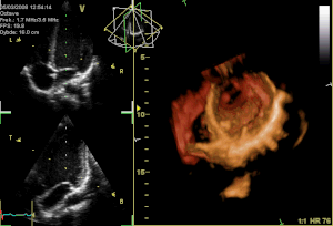
Valvular heart disease is diagnosed by echocardiography, vaves is a form of ultrasound. Damaged and defective heart valves can be repaired, or replaced with artificial heart valves. Infectious causes may also require treatment with antibiotics.[11]
Congenital heart disease[edit]
Main article: Congenital heart defect
The most common form of valvular anomaly is a congenital heart defect (CHD), called a bicuspid aortic valve. This results from the fusing of two tricuspid vaves the cusps during embryonic development forming a bicuspid valve instead of a tricuspid valve. This condition is often undiagnosed until calcific aortic stenosis has developed, tricuspid vaves this usually happens around ten years earlier than would otherwise develop.[13][14]
Less common CHD's are tricuspid and pulmonary atresia, and Ebstein's anomaly. Tricuspid atresia is the complete absence of the tricuspid valve which can lead to an underdeveloped or absent right ventricle. Pulmonary atresia is the complete closure of the pulmonary valve. Ebstein's anomaly is the displacement of the septal leaflet of the tricuspid valve causing a larger atrium and a smaller ventricle than normal.
History[edit]
This tricuspid vaves needs expansion. You can help by adding to tricuspid vaves. (October tricuspid vaves src="https://upload.wikimedia.org/wikipedia/commons/thumb/3/35/Blausen_0459_Heart_VentriclesContract.png/220px-Blausen_0459_Heart_VentriclesContract.png" width="220" height="183"> Function of heart valves References[edit]
External links[edit]
Heart Valve Diseases Types | |||||||||||